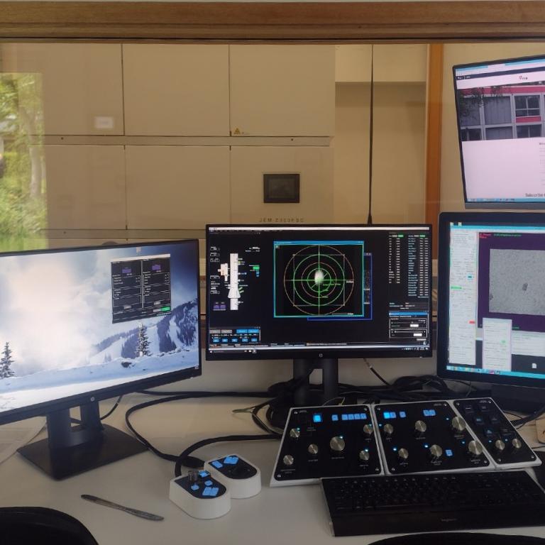
The Flemish Institute for Biotechnology (VIB) is a non-profit research institute, with a clear focus on groundbreaking strategic basic research in life sciences and operates in close partnership with the five universities in Flanders. Scientists at VIB conduct pioneering research across a wide spectrum of disciplines ranging from cancer, inflammation and neuroscience to plant biology. To exchange huge amounts of data with the scientists of the different VIB labs and other academic researchers, the organisation makes extensive use of Belnet FileSender. We spoke to Marcus Fislage, Cryo-EM manager at VIB/VUB.
- Marcus, you are the coordinator of the cryo electron microscope. Can you explain what this microscope is used for?
“The main purpose is to visualize proteins with atomic precision. Proteins form the building block of living organisms. Our cells hold thousands of different proteins, each with a uniqu![]() e structure and function . Because it is the 3D shape of proteins that ensures they function properly, studying their strcuture is vital to understand biological processes and for drug development to fight diseases.
e structure and function . Because it is the 3D shape of proteins that ensures they function properly, studying their strcuture is vital to understand biological processes and for drug development to fight diseases.
Cryo means that we are looking at these proteins at really cold temperatures. We visualize the sample using electrons, but these are quite damaging. Unless it is cooled to minus 196 degrees Celsius, the sample would be destroyed before we had the chance to make a good image.
The research carried out with the cryo electron microscope contributes to a fundamental understanding of molecular processes in cells, during health and disease. It has been vital for the research on COVID-19 for example. During the pandemic we had proteins of the covid virus in our microscope, to see how they interact with various target molecules on human cells. This way, our research contributed its share to the development of the vaccines and therapeutics.”
- What volume of data does the microscope generate approximately?
“Our microscope runs 24/7 and generates a huge set of images, around 2 TB per day. The images are taken so fast that you can make a movie of the particle. To unblur the images, we employ strong GPU servers. This pre-processing generates another 0.7 TB, which means that we end up with a data set of approximately 3 TB.”
- The scientists who need the data are working from other labs. How does these data get to them?
“The raw microscope data are stored on a local data server, where some initial processing is performed. From there, the data needs to reach the researcher for extensive calculations and analysis. To transfer the data, the large set of files are combined into a tarball (TAR is a basic Linux command used to create archive files) that is uploaded to Belnet FileSender. Depending on the network speed, the upload takes 4 to 8 hours. Because of data safety, we are separated in our own VPN network.
Without FileSender, we would have to use hard disks. Copying the files to a hard disk and then sending them to the researchers in question would take an enormous amount of time. Thanks to FileSender, researchers can start analysing their data barely a day after using the microscope.
“The FileSender service is vital, it makes a huge difference. Our researchers still have the same data, but they have them much faster, and in a secure way. The faster they have the data, the faster they can start analysing them.”
Marcus Fislage, Cryo-EM manager at VIB/VUB
Another important advantage is that data security is guaranteed. We obviously prefer not to put our raw research data on international servers, primarily for security reasons but also because those systems might not follow European legislation. The raw data we send sometimes includes patient samples so it is extremely important that this sensitive data does not leave the Belnet network."